


Fluoro tumour finder tracking well
 Print
Print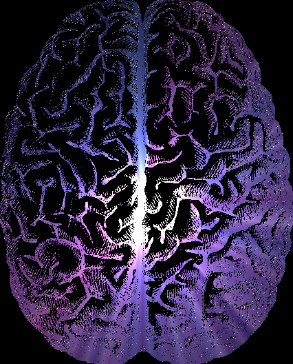 Tests have shed light on new ways to find brain tumours, by making them much easier to spot.
A recent study looked at the use of two special compounds that contain fluorescent dyes which are attracted to cancer cells.
Researchers in the US evaluated two “tumour-selective” fluorescent agents - called CLR1501 and CLR1502 - for their ability to differentiate brain tumours from normal brain tissue in mice.
CLR1501 and CLR1502 are synthetic analogues of the tumour-targeting agent alkylphosphocholine (APC), which is specifically attracted to cancer cells.
The synthetic form was molecularly altered to carry fluorescent dyes that glow under lights with specific wavelengths in the green (CLR1501) or near-infrared (CLR1502) range.
Viewed under appropriate conditions, the dyes did indeed make tumour cells ‘light up’, so they could be readily distinguished from normal neighbouring brain tissue.
The near-infrared imaging with CLR1502 successfully localised brain tumours through the intact skin and skull of living mice.
Both APC analogues provided “excellent fluorescence discrimination of tumour from adjacent normal brain,” the report says.
The tumours could be clearly spotted using a number of commercially available imaging systems.
When used during surgery for brain cancer, the team behind the project at the University of Wisconsin School of Medicine and Public Health say the fluorescent dyes could help neurosurgeons to locate a tumour and to resect it as completely as possible.
Removing all visible areas of cancer (gross total resection) significantly improves survival after brain cancer surgery.
Outside of neurosurgery, researchers hope similar APC analogues might be useful in diagnosing brain tumours, as well as targeting chemotherapy drugs directly to cancer cells.
University of Wisconsin’s Dr John Kuo and co-authors write; “Upcoming clinical trials in human tumours are planned for these promising tumour-selective fluorescence agents.”