


Advanced scanner gives edge on cancer
 Print
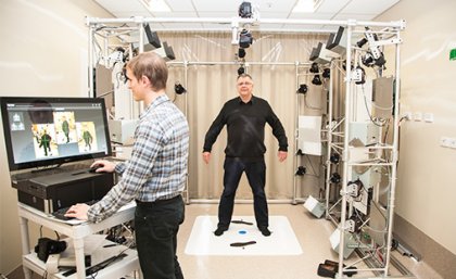
Print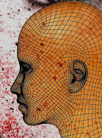 A high-tech rig containing 46 cameras is Queensland’s newest weapon in the fight against skin cancer.
In the state with some of the highest melanoma rates in the world, researchers now have access to technology that will improve diagnoses and could save lives.
The University of Queensland has fired up its new VECTRA Whole Body scanner.
The device makes 3D maps of patients, which are used to spot and monitor the precursors to skin cancer.
“The primary use of the total body photography system is tracking changes in skin lesions which are a tell-tale sign of a developing melanoma,” says Professor Peter Soyer, director of UQ’s Dermatology Research Centre.
“It can also be used to measure body dimensions and track these over time.”
To make a 3D avatar, a patient stands within a scaffold supporting 46 cameras, which each take an image at the same time.
A computer program stitches the images together to produce a 3D model that replicates the skin’s surface in fine detail.
A separate camera can be used to make dermoscopic images of individual lesions, linked to their location on the 3D model.
The prototype, funded by the Private Practice Trust Fund, is the first of its kind outside New York, where there are two at the Memorial Sloan Kettering Cancer Center.
 Research assistant Mr Glen Wimberley said the response from participants so farhad been positive.
“Patients really like the system,” Mr Wimberley said.
“They are fascinated by the technology and can see the potential benefits.”
The VECTRA Whole Body 360 technology is in the Clinical Research Facility of the Translational Research Institute at the Princess Alexandra Hospital.