


Ultrasound drug delivery tested
 Print
Print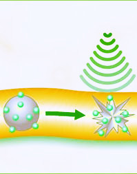 A trial in the UK has tested a new way to deliver chemotherapy driven by ultrasound.
The technique involves heat-sensitive lipid capsules filled with chemotherapy drugs, which are ruptured remotely using ultrasound.
The phase 1 clinical trial testing found that the technique increased the amount of chemotherapy delivered directly to liver tumours.
“Reaching therapeutic levels of cancer drugs within a tumour, while avoiding side effects for the rest of the body is a challenge for all cancer drugs, including small molecules, antibodies and viruses,” says Professor Constantin Coussios from the University of Oxford.
“Our study is the first to trial this new technique in humans, and finds that it is possible to safely trigger and target the delivery of chemotherapy deep within the body using focussed ultrasound.
“Once inside the tumour, the lipid capsules release the drug, supplying a higher dose of chemotherapy directly to the tumour, which may help to treat tumours more effectively.”
The open-label phase 1 trial was conducted in one hospital in the UK, and included 10 patients with incurable tumours in their livers – including cancers of the liver, as well as tumours that had spread to the liver from other areas of the body.
The technique is completed under general anaesthetic. A dose of 50mg/m2 of doxorubicin chemotherapy within heat-sensitive lipid capsules is injected into each patient’s bloodstream.
A beam of ultrasound was then focussed on the liver tumour, slightly increasing the temperature in that area. Above 39.5⁰C, the capsules release the drug into the bloodstream inside the tumour.
This then creates a high drug concentration gradient, driving more chemotherapy into the tumour.
The researchers measured how much ultrasound exposure was needed to heat the tumour to 39.5⁰C or higher by temporarily implanting a heat-recording device placed into six patients’ tumours. They also took tumour biopsies from all 10 patients to assess the amount of chemotherapy delivered inside the tumour.
In seven out of 10 patients, chemotherapy concentrations within the liver tumour were two or more times higher after the ultrasound, compared with before ultrasound exposure, when there was only passive build-up of chemotherapy from the liposomes in the tumour.
The concentration of chemotherapy within the tumour was less than doubled in the other three patients.
On average, the new technique increased the amount of chemotherapy inside the tumour by 3.7 times (from 2.34 µg/g chemotherapy within the tumour before ultrasound exposure, to 8.56 µg/g chemotherapy within the tumour after ultrasound exposure).