


Biobank to spread virtual cancer
 Print
Print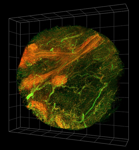 Australian researchers have developed the world’s first virtual platform to host 3D copies of human cancer tissues.
The Virtual Biobank will digitise be used to tissue samples donated by patients, which up until now could only be requested through physical biobanks.
Each digital cancer sample in The Virtual Biobank is made up of high resolution microscopy images in both 2D and 3D, plus important clinical and molecular information.
Researchers have already taken 20 breast cancer samples from tumour biopsies stored at the Hunter Cancer Biobank and converted them into virtual copies, enabling anyone with an internet connection to carry out research from their computers.
“It currently takes many months before researchers are able to obtain tissue samples from a physical biobank and carry out investigations with it. Once a researcher has performed their study, that sample typically cannot be reused,” says lead investigator on the biobank project, Dr Jamie Flynn.
“This process ensures the physical sample remains intact, but a 3D, digital copy with clinical and experimental information is kept online for future use.
“This is particularly critical for rare cancers, which are hard to study due to a limited number of samples.”
Hunter Cancer Biobank Scientific Advisor, Dr Rick Thorne, said The Virtual Biobank is key to unlocking the third dimension of cancer tissues.
“Putting these samples in their true context for the first time will help researchers with many different problems, including how to get treatments to all of the cancer cells in tumours,” Dr Thorne said.
“The project has kicked off with breast cancer, but the processes used by the team are applicable to any cancer tissue. It is remarkable this will happen online and we look forward to future expansions of the dataset.”
The team embedded specialised software tools into the online platform that would otherwise be too expensive for researchers to access.
“We really wanted to make The Virtual Biobank interactive and dynamic, so with lead developer, Dr Bill Pascoe … we developed a software toolkit for other people to contribute additional information such as findings from their own investigations,” Dr Antony Martin said.
The ‘Tissue Tracker’ component of the site allows users to automatically log the experimental procedures for all new tissue samples coming into a lab, removing the need for hand-written notes and allowing other researchers to easily replicate experiments.
“Users can also create full 3D images for free by simply uploading their own microscopy data to our ‘Terastitcher Pipeline’, eliminating the need for expensive commercial software that can ordinarily cost upwards of $50,000,” Dr Martin said.
While still in its early stages, the online platform holds promising future potential in areas of research, education and even virtual reality.
“The technology and tools that we’ve put together make this data available to anyone in the world. People in the fields of engineering or computer science can now easily apply their knowledge to questions surrounding cancer,” Dr William Palmer said.
“Educators can also access the site and use real-world examples to teach pathology to the next generation of cancer clinicians and researchers.
“We’d also like to convert the 3D data into virtual reality for education and general awareness. Hopefully soon, anyone with a smartphone and Google Cardboard could experience the internal environment of cancer tissue and bring about new insights.”