


Low lights bring closer view
 Print
Print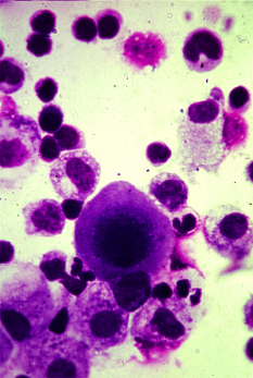 Australian researchers have found a simple way to unlock the nanoscale world on standard biology lab equipment.
Standard optical microscopes can image cells and bacteria but not their nanoscale features which are blurred by a physical effect called diffraction.
Optical microscopes have evolved over the last two decades to overcome this diffraction limit; however, these so-called super-resolution techniques typically require expensive and elaborated instrumentation or imaging procedures.
Now, Australian researchers say they have found a simple way to bypass diffraction limitations.
“We have identified a particular type of fluorescent markers, so-called ‘upconversion nanoparticles’, that can enter into a regime in which light emitted from the particles grows abruptly – in a super-linear fashion – when increasing the excitation light intensity,” says Dr Martin Ploschner from the ARC Centre of Excellence for Nanoscale BioPhotonics (CNBP).
“Our key discovery is that if this effect is exploited under the right imaging conditions, any standard scanning optical microscope can spontaneously image with super-resolution.
“While we have chosen to demonstrate this upconversion super-linear excitation-emission (uSEE) on one of the most commonly used types of optical microscopes – a confocal microscope – practically any type of scanning microscope or microscope involving variations in the illumination intensity can benefit from this spontaneous improvement of the resolution.”
Fellow researcher Dr Denitza Denkova says the uSEE approach improves the resolution beyond the diffraction limit simply by reducing the illumination intensity.
“Our approach works in the opposite direction to all other existing super-resolution methods; the lower the laser power, the better the resolution and the lower the risk of photo-damage to the bio-samples,” she says.
“Best of all, super-resolution can be achieved without setup modifications and image processing. Thus, this method has the potential to enter any biological lab, at practically no extra cost.
“The value of our work is in realising the technique, for the first time, in a 3D biological setting, using biologically convenient particles.
“We suggest a modification of the composition of the nanoparticles and the imaging conditions, which triggers the spontaneous super-resolution to occur under a practically relevant microscopy configuration.”
The research team has also developed a theoretical framework which allows end-users to adjust the particle composition and the imaging conditions and achieve super-resolution in their own laboratory setting.
“Our work enables microscopists to look in a new way with their existing tools,” she said.