


Cell-stripping for naked matrix
 Print
Print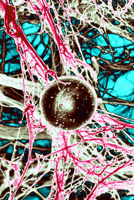 Researchers have unveiled a new technique for stripping the cells out of the extracellular matrix surrounding them.
The extracellular matrix has a profound impact on how cells behave and is important in the progression and spread of cancer.
The new technique dissolves cells away from tissues, leaving behind an unaltered matrix that can be viewed – and its components catalogued – in unprecedented detail in 3D.
“We have developed a technique to obtain intact organ scaffolds and to image them in incredibly high detail using microscopes. We are the first to image the 3D structures of primary and metastatic tumours as well as healthy organs in this way,” says Professor Janine Erler from the University of Copenhagen.
Developed in mice post mortem, the technique is the first to use existing blood vessels to deliver cell-removing compounds directly to a specific tissue or organ.
“We have isolated the structure that keeps tissues in place and organises the cells inside them. We did this by using existing blood vessels to deliver cell-removing compounds directly to a specific tissue to remove all cells within an organ,” says postdoctoral fellow Dr Alejandro Mayorca-Guiliani.
“Doing this leaves behind an intact scaffold that could be analysed biochemically and microscopically, providing us with the first view of the structure of tumours.”
Imaging expert and co-first author Dr Chris Madsen says; “When you remove the cells, the clarity of what you can see through the microscope is much improved – you can see the fibres of the matrix more clearly and you can look much deeper into the tissue”.
“Using this approach, we have been able to see important differences in matrix organisation when we looked at metastatic tumours in the lung and in the lymph node,” he said
Matrix biology and mass spectrometry expert and co-first author Dr Thomas Cox (now based at the Garvan Institute of Medical Research, Sydney) says; “Because we are removing the cells completely, we can use mass spectrometry to identify and catalogue the components of the matrix – in normal tissue and in tumours – in unprecedented detail”.
“What is really exciting is we found that some of the components of the matrix in different secondary tumours [metastases] are unique to that tissue. That is telling us that remodelling of the matrix in cancer is organ-specific.”
By using the decellularised organs, experts can learn much more about how tumours and normal organs are built, and what their differences are. This new technique might even have an impact on organ regeneration and tissue engineering in the future.
“We are now re-introducing cells into our extracellular matrix scaffolds, bringing them back to life, to study how tumours form and how cancer progresses. This is extremely exciting and offers a unique opportunity to study how cells behave in their native environment,” Professor Erler explains.